Descripción
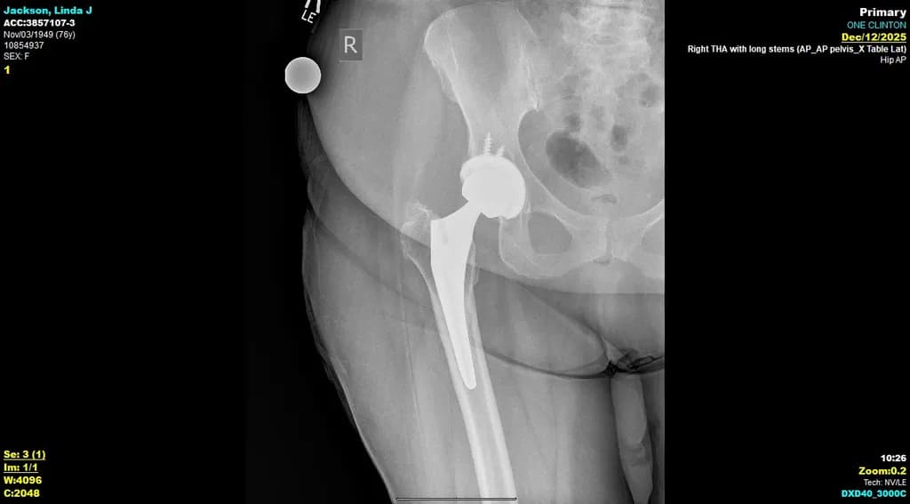
This X-ray image reveals a right total hip arthroplasty featuring long stems, clearly presenting the alignment and integration of the prosthetic components within the pelvic bone. The contrast highlights the precise placement and stability of the implant against the natural bone structure. An informative visual for medical professionals or educational settings, it contributes valuable insight into orthopedic procedures. This piece would be a distinctive addition to any clinical or academic environment.